Introduction
In a female of reproductive age group, clinical presentation as a cystic mass in the abdomen poses a diagnostic dilemma. With the advent of imaging techniques like CT scans and MRI, accurate diagnosis can be made in these cases. The commonest diagnosis for a cystic mass in the pelvis is ovarian cyst. However extensive cystic degeneration of fibroids, especially in the broad ligament can mimic a ovarian cyst.1 When the leiomyoma increases in size, the vascular supply to it becomes inadequate and leads to different types of degeneration: hyaline, cystic, myxoid, or red degeneration. Cystic degeneration is an uncommon type of degeneration that a uterine leiomyoma (fibroid) can undergo. This type of degeneration is thought to represent ~4% of all types of uterine leiomyoma degeneration.1 Here we report a case of cystic degeneration of broad ligament fibroid mimicking as a ovarian cyst.
Case Report
A 45year old female was referred to our OPD as a case of ovarian cyst. She presented with complaints of abdominal discomfort for past 3months which was gradual in onset and progressive in nature and was associated with dull aching pain and heaviness in the lower abdomen. There was no history of menstrual irregularities, loss of weight or appetite. The patient has 2 living issues with normal vaginal deliveries.
On examination patient general condition was fair, afebrile, vitals stable, no pallor, no pedal edema. Per abdominal examination revealed a pelvic mass corresponding to 20 weeks gravid uterus, regular margins, cystic in consistency, mobile, non-tender lower border palpable. Speculum examination revealed cervix and vagina healthy. Bimanual pelvic examination showed uterus corresponding to 20 week gravid size, mobile, cystic and non-tender and the mass moves with cervical movement. Left and posterior fornices were full, no tenderness.
Investigations
Her USG findings are Right adnexal heteroechoic mass of size 12 x 7.8 x 13.7 cm with few internal cystic spaces noted within. No evidence of vascularity noted. Right ovary not seen separately. Suggestive of broad ligament fibroid with differential diagnosis of ovarian mass. MRI pelvis showed a large heterogeneous lesion of 14cm × 7.7 cm × 4.4 cm with irregular contour and internal cystic areas in the pelvis with extension to the abdominal region. The right ovary is not separately visualized. Uterus is grossly displaced to the left by the mass, posterior wall of the uterus appears bulky, and asymmetrical and shows seedling fibroid. No evidence of ascites and lymphadenopathy. Her routine blood investigations and serum biochemistry were within normal limits. CA 125 was also normal. With the working diagnosis of broad ligament fibroid and differential diagnosis of ovarian mass, the patient was taken up for laparotomy.
Intra-operative findings
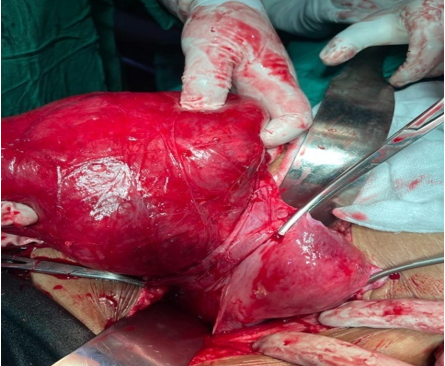
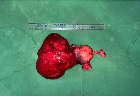
The intra-operative diagnosis was huge broad ligament fibroid. Intra-operatively, following a midline longitudinal incision with extension over the umbilicus, a 17 × 15 cm sized fibroid was noted in the Right broad ligament (Figure 1). Uterus was pushed to the left side. Uterus was normal, left fallopian tube and round ligament looked grossly normal, and both ovaries were normal. Careful dissection was done to prevent ureteric injuries. Excision of tumor with total abdominal hysterectomy and bilateral salpingo-oopheretomy was done (Figure 2). It was a false broad ligament fibroid which has undergone cystic degeneration.
Her postoperative period was uneventful and was discharged. The patient is on follow up.
Histopathology
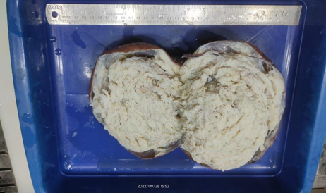
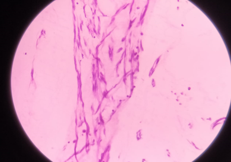
On gross examination, a single large globular soft tissue mass measuring 20x16x7cms was found. Cut section of the tumour showed a grey white mass with multiple cystic areas filled with a mucoid and gelatinous material.(Figure 3) Microscopic examination showed benign leiomyoma with cystic and focal myxoid degeneration.(Figure 4)
Discussion
Broad ligament is a two layered fold of peritoneum connecting the sides of uterus to lateral walls of pelvis and its floor. Broad ligament leiomyoma can originate from the uterus and invade the broad ligament (false) or it can originate from broad ligament itself (true). These benign tumors are usually asymptomatic. However, if the leiomyoma reaches significant size, it can potentially compress the surrounding pelvic structures and manifest clinically with various signs and symptoms.1
A leiomyoma or fibroid is a benign tumour with a prevalence of 20%-30% in the women of reproductive age and more than 40% of women above 40 years of age.2 If allowed to reach an enormous size, these broad ligament fibroids can present with pressure symptoms, pelvic pain, bladder and bowel dysfunction. Intrauterine fibroid on the other hand in addition to pressure symptoms often presents with menstrual abnormalities and dysmenorrhea.
The case reported here presented only with abdominal discomfort without any menstrual abnormalities, likely to have been caused by the huge broad ligament. The differential diagnosis for broad ligament leiomyoma includes ovarian masses (benign or malignant), tubo-ovarian masses, broad ligament cyst and lymphadenopathy. In our case on clinical examination and ultrasonography it was suspicious of an ovarian tumor or broad ligament fibroid. The serum level of cancer antigen CA-125 was done which was in normal range.
It is important for any adnexal masses to discriminate between benign and malignant nature of the lesion in the preoperative period for optimal patient management.2
Leiomyomas may be single or multiple. In our case, there was a single mass in broad ligament. Broad ligament leiomyomas have the potential to grow to large size.3 When they grow to a large size, secondary changes may occur which includes degeneration, infection, hemorrhage, and necrosis. The cystic changes in lesion mimic the metastatic malignant ovarian tumors.4, 5 It is important for any adnexal masses to discriminate between benign and malignant nature of the lesion in the preoperative period for optimal patient management.6, 7
Uterine leiomyoma usually does not pose clinical and sonological diagnostic challenges. Radiological investigations are often requested for to assess the extent of the mass and its relationship with other pelvic and abdominal structures. However, extra uterine fibroid distorting the pelvic anatomy may raise suspicion of an ovarian tumour.8
Conclusion
Benign tumors in the broad ligament are usually asymptomatic but if left untreated for a long time, it can reach an enormous size resulting in chronic pelvic pain, compression of adjacent structures. They can also undergo cystic degeneration posing difficulty in diagnosis. These tumours with cystic degeneration can mimic ovarian cyst on imaging as the normal contour of fibroid is lost. Ovarian malignancy should be considered prior to a planned surgery.


