- Visibility 58 Views
- Downloads 4 Downloads
- DOI 10.18231/j.sajcrr.2023.007
-
CrossMark
- Citation
Unusually large number of gall stones removed through laparoscopic cholecystectomy from a single patient- A case report
Introduction
Gallstone disease remains one of the most common medical problems leading to surgical intervention.[1] The incidence of gallstones increases with age, with females more likely to form gallstones than males.[2] Epigastric and right upper quadrant pain occurring 30-60 minutes after meals is a common presentation and ultrasonography is the diagnostic test of choice.[1] Laparoscopic cholecystectomy (LC) since its inception in 1989 has become the gold standard treatment for gall stone disease.[3] It has been linked to a lower complication rate and shorter postoperative hospital stay as compared to open cholecystectomy.[4]
In the present case report, we describe the case of a 43-year-old woman who presented with right hypochondriac pain and a subsequent laparoscopic cholecystectomy involved removal of an extremely large number of gall stones.
Case Report
A 43-year-old lady presented to the Surgical Outpatient Department in August 2022, with the chief complaint of on and of pain in the right hypochondriac region since December 2021.
An ultrasound (USG) of the abdomen was done on the 7th of December for the same, which showed a hugely distended gall bladder (12.3 cm in length) with multiple tiny calculi, including a 6mm calculus at the neck. This was followed by a three tesla MR cholangiopancreatography on 12th December 2021, which showed a markedly distended gall bladder with presence of multiple faceted signal void area representing calculi in the fundus, body and neck region. The largest stone measured 19x14mm and there was no evidence of cholecystitis or choledocholithiasis.
She also complained of associated nausea and occasional episodes of vomiting. The patient had no other co-morbidities.
On examination, the abdomen was soft and revealed no tenderness or palpable mass. The rest of the examination was unremarkable.
A USG of the abdomen and pelvis was done on 17th of August 2022, which showed an over-distended gall bladder. Multiple gall bladder calculi were noted, the largest being 3.7mm in size. This was consistent with the results of an earlier USG done on 21st March 2022.
Blood investigations including liver function tests were within normal limits.
Thereafter, the patient was admitted for surgical management. Informed consent was obtained after explanation of the surgical procedure and the possible complications.
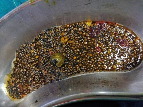
A laparoscopic cholecystectomy was done on 22nd August under general anesthesia. Four small incisions were made in the patient’s abdomen and a tube with a tiny video camera was inserted into the abdomen through one of the incisions. The gall bladder was removed using surgical tools inserted through one of the incisions, under the guidance of a video monitor to ensure accuracy. A total of seven thousand two hundred and twenty-eight stones were removed ([Figure 1]). The procedure itself took about 40 minutes while counting the stones took around 4 hours. As per the best of our knowledge, this is among the largest reported number of stones removed via a laparoscopic procedure. There were no intra or post-operative complications and the patient was discharged on 24th of August 2022.

Discussion
Cholelithiasis or gallstone disease involves formation of concretions in the gall bladder. An estimated 20% of adults over 40 years of age and 30% of over 70 years of age have biliary calculi. The risk factors include obesity, diabetes mellitus, oestrogen and pregnancy, haemolytic diseases, and liver cirrhosis.[1] 75% of gallstones are composed of cholesterol, and the other 25% are pigmented. Despite the varied composition of gallstones, the clinical signs and symptoms are the same.[2] The presence of gallstone disease is best described as a continuum from asymptomatic to symptomatic disease, with the latter including both pain attacks and complicated disease.[3] The pain (biliary colic) is characteristically steady, usually moderate to severe in intensity, located in the epigastrium or right upper quadrant of the abdomen and is caused by the intermittent obstruction of the cystic duct by a stone. [3], [4] If pain persists with the onset of fever or high white blood cell count, it should raise suspicion for complications such as acute cholecystitis, gallstone pancreatitis, and ascending cholangitis.[4] Expectant management is considered the most appropriate choice in patients with asymptomatic gallstones.[5] Symptomatic disease causes a persistent high risk of symptom recurrence and need of cholecystectomy.[3]
The diagnosis of chronic cholecystitis is made by the presence of biliary colic with evidence of gallstones on an imaging study. Ultrasonography being 90-95% sensitive, is considered the test of choice although additional imaging studies may be indicated.[1], [4] Laparoscopic cholecystectomy, a minimally invasive surgical procedure, has essentially replaced the open technique for routine cholecystectomies.[2] It allows shorter hospitalization, rapid recovery and early return to work.[6] The standard technique of performing LC is to use 4 ports.[7]
The most significant common complication is injury to the bile duct.[8] However, injuries during the procedure can be prevented by precise operative technique, clear visualisation of anatomical landmarks, and careful dissection of tissues. Minor complications are usually treated conservatively. Major complications (biliary and vascular) are life threatening and increase mortality rate, hence creating the need for conversion to an open surgical approach in order to treat them.[9]
The risk of postoperative complications is increased by risk factors like male gender, high age, impaired renal function and conversion to open surgery.[10]
Conclusion
The relevance of this case is due to the large number (7,228) of gall stones removed via laparoscopic route of surgery without any complications. This is among the highest number of gall stones removed laparoscopic ally in India.
Source of Funding
None.
Conflict of Interest
None.
References
- BD Schirmer, KL Winters, R Edlich. Cholelithiasis and cholecystitis. J Long-term Effects Med Imp 2005. [Google Scholar]
- KR Hassler, JT Collins, K Philip, MW Jones. . Laparoscopic cholecystectomy 2022. [Google Scholar]
- DM Shabanzadeh. Incidence of gallstone disease and complications. Curr Opin Gastroenterol 2018. [Google Scholar]
- S Abraham, HG Rivero, IV Erlikh, LF Griffith, VK Kondamudi. Surgical and nonsurgical management of gallstones. Am Fam Physician 2014. [Google Scholar]
- KS Yoo. Management of gallstone. Korean J Gastroenterol 2018. [Google Scholar]
- GP Sadler, A Shandall, BI Rees. Laparoscopic cholecystectomy. Brit J Hospital Med 1992. [Google Scholar]
- SP Haribhakti, JH Mistry. Techniques of laparoscopic cholecystectomy: Nomenclature and selection. J Minim Access Surg 2015. [Google Scholar]
- VS Lee, RS Chari, G Cucchiaro, WC Meyers. Complications of laparoscopic cholecystectomy. AM J Surg 1993. [Google Scholar]
- M Radunovic, R Lazovic, N Popovic, M Magdelinic, M Bulajic, L Radunovic. Complications of Laparoscopic Cholecystectomy: Our Experience from a Retrospective Analysis. Open Access Maced J Med Sci 2009. [Google Scholar]
- PM Terho, AK Leppäniemi, PJ Mentula. Laparoscopic cholecystectomy for acute calculous cholecystitis: a retrospective study assessing risk factors for conversion and complications.. World J Emerg Surg 2016. [Google Scholar]
