- Visibility 62 Views
- Downloads 47 Downloads
- DOI 10.18231/j.sajcrr.2024.022
-
CrossMark
- Citation
Chondroblastoma of scapula with aneurysmal bone cyst, unusual presentation at unusual location
Introduction
Chondroblastoma is a rare benign bone tumor of cartilage origin which was first reported as “epiphyseal chondromatous giant cell tumor” by Codman in 1931. Later chondroblastoma as separate entity given by Jaffe and Lichtenstein in 1942.[1] About 1% of all benign bone tumours are chondroblastoma, which are most commonly diagnosed in young male patients and reach their peak occurrence in the second decade of life. Typical tumor locations are the long bones epiphysis femur, tibia and proximal humerus. Only 16% of cases are found in flat bones, pelvic bones are the most frequently involved, followed by the calcaneus, pelvis, ribs and scapula. Chondroblastoma of scapula is extremely rare and among that, around 20% chondroblastoma associated with aneurysmal bone cyst.[2]
Case Presentation
A 65-year female presented with pain and limitation of right shoulder mobility. Physical examination shows a tumor at the inferior angle of right scapula with atrophy of the right shoulder girdle musculature compared to the left side. Active adduction/abduction accounted for only 30–0–20, the passive one for 30–0–40 degrees with a restriction of rotational movement. There were no lymphadenopathy, fever, chills, or weight changes and all the laboratory findings were within normal ranges.
Xray: Well defined lucent lesion with thin sclerotic rim at inferior angle of scapula.
Computed Tomography showed a well demarcated expansile lytic lesion measuring 51x31mm in size with lobulated margins seen arising from the inferior angle of the right scapula. Thinning of the cortex seen at few places.
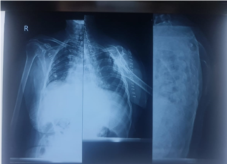
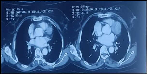
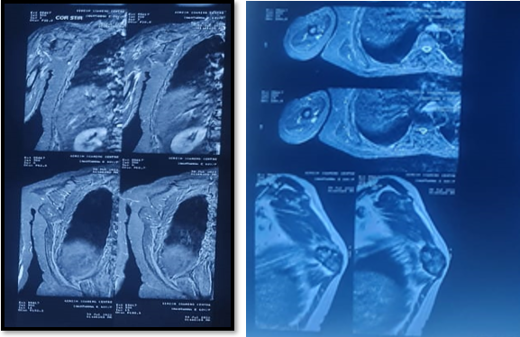
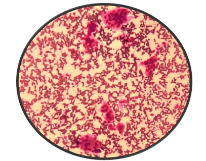
MRI right scapular showed that exophytic T2 hypointense & STIR mixed intensity lesion measuring 5 x 4.5x2.3cms in inferior angle of scapula suggesting Benign exophytic lesion from inferior angle of scapula with surrounding soft tissue mass Exostosis.
FNAC of right scapular lesion showed in dense haemorrhagic background multinucleated giant cells containing 20-80 nuclei which are round to oval uniform with bland chromatin and inconspicuous nucleoli. The giant cells have moderate amount of amphophilic cytoplasm well defined border. Also seen are scattered small groups of dispersed plumps to fusiform cells with round to oval uniform multinucleated giant cell nuclei. No malignant cells. A diagnosis of Giant cell tumour of bone with differential diagnosis Aneurysm bone cyst / Chondroblastoma suggested.
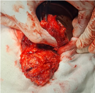
Excision of right scapular lesion done under the general anaesthesia and tumour specimen sent for histopathological examination.
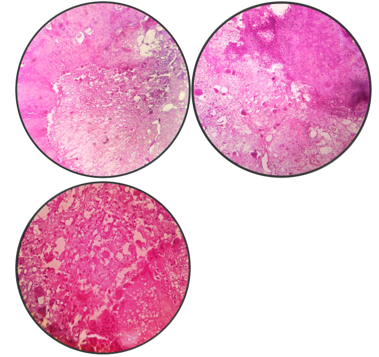
Histopathological findings of the surgical specimen revealed that a solid and cystic tumour composed of mixture of sheets of mononuclear cells and multinucleated giant cells. These mononuclear cells are round to polygonal with clear cytoplasm and distinct cytoplasmic birders. The nucleoli are oval bland with few longitudinal groves and occasional mitosis. Irregular zone pf focal calcification (chicken wire type and areas of chondroid differentiated noted. Interspersed among these are multinucleated osteoclastic type giant cells. Also seen are large areas of haemorrhage rimmed by osteoclast giant cells noted (ABC like areas). No evidence of malignancy. Histological features are of Chondroblastoma with secondary Aneurysmal bone cyst.
Post operative course were uneventful.
Discussion
Chondroblastoma was originally classified with the giant cell tumors because presence of osteoclast-like giant cells by Ewing in 1928. The histopathological classification was revised by Codman in 1931 to "epiphyseal chondromatous giant cell tumor". Benign chondroblastoma was finally identified as a distinct tumor in 1942 by Jaffe and Lichtenstein. Chondroblastoma is rare, accounting for approximately 1% of all benign bone tumors. Most cases occur during the 2nd decade of life. Especially in older patients, there were reported a few atypical cases of chondroblastoma, which tend to involve uncommon sites and have a tendency to expand the affected bone. The presenting symptoms are mild pain and, less often, local swelling associated with limitation of motion of the adjacent joints. The long bones epiphysis of the femur, humerus, tibia and tarsal bones are the most common location for chondroblastoma. Chondroblastoma is usually benign, but malignant lesions have been reported. The benign nature of most chondroblastoma is demonstrated by the 90% cure rate from simple curettage. [3]
Chondroblastoma's histogenesis is debatable, however epiphyseal cartilage cells rather than reticulo-histiocytic cells are where it originates. The histological diagnosis of typical chondroblastoma is usually easy due to their characteristic appearance of rounded or eosinophilic chondroid, multinucleated giant cells, or polygonal chondroblasts, extracellular matrix with focal (chicken wire) calcification. [2]
In radiologically scalloping or expansion of cortical bone with calcifications, either punctate or in rings, may be visible. In MRI and CT 20% cases associated with cyst. The proximity of the tumour to the joint, the integrity of the cortex, and intralesional calcifications can all be determined with a CT scan. When the patient's age and the lesion's location, which are diagnostic of the lesion, are visible on radiographs, chondroblastoma can be diagnosed. Radiographic diagnosis of an atypical chondroblastoma is more difficult because of many differential diagnoses, including benign and malignant lesions in the area of predilection. The above-mentioned imaging features were highly suggestive of a benign, cystic but locally invasive process can be present. The differential diagnosis included Aneurysmal bone cyst, chondroblastic osteosarcoma, chondrosarcoma and fibrous dysplasia. Generally, ABCs present with pain and swelling with related to compression of other surrounding structure. Since the periosteum is intact around the soft tissue component in ABC, the characteristic exhibits more precisely defined edges and peripheral egg shell calcification (a benign radiographic feature). Intralesional fluid–fluid levels are common to both chondroblastoma and ABCs and are, therefore, not generally helpful for distinguishing the two entities. To make this judgement, a histological study is necessary. Benign chondroblastoma with ABCs is often treated with intralesional curettage excision and bone grafting. In most instances, removing the tumour is sufficient. The scapular placement of the tumour, the difficulties in making a diagnosis, and the available treatments all seem to make the current case special in this situation. [4]
According to the latest World Health Organization classification 2016, CB is classified as an intermediate tumor that is locally aggressive and rarely metastasizes (< 2%), with a local recurrence rate of up to 38% and a 2% incidence of lung metastases. In general, local recurrence is more likely to occur in flat than in long bones.1 The recurrence rate of chondroblastoma after complete excision was reported to be approximately 20%, and approximately 50% in patients who underwent curettage. The rate of recurrence was approximately two-fold higher than that in patients treated with complete excision and curettage. Therefore, complete resection is dependent on the extent of the tumor is essential for postoperative local control. Therefore, strict follow-ups are needed due to the possibility of local recurrence. [5]
The need for a combined clinical, radiological, and histopathological approach to the correct diagnosis of benign chondroblastoma with secondary ABC is emphasised. Chondroblastoma of flat bone is a very rare condition as shown in the present case, most of the classic criteria such as typical location and radiological appearance were not seen in the present case. Chondroblastoma rarely exhibits malignant behaviour and metastatic spread. It can be difficult to diagnose a chondroblastoma in the scapula. Some tumours may manifest in a unique way. As was the situation with this patient, not all traditional radiological findings are always present. As a result, a histological study of the tumour is always necessary. [4]
Conclusion
The clinical, radiological and histopathogical findings are necessary for evaluation of chondroblastoma and to avoid erroneous diagnostic conclusions. Chondroblastoma does occasionally metastatically spread and exhibit malignant behaviour, however this is extremely uncommon. Complete excision and curettage of the tumour with a regular follow-up for any local recurrence or metastasis is the key to successful management of such case. The histopathological diagnosis should not be stopped at chondroblastoma a careful search for associated aneurysmal bone cyst is essential, as most cases of chondroblastoma associated with this lesion.
Authors’ Contributions
We Dr. Sainath k Adola, Dr. Soneji Dhairya Kirtikumar and Dr. Nishant Panegar doing this case report. Dr. Nishant did operative proccedure like excision of tumour of inferor angle of scapula after that specimen sent for histological examination, in histopathological diagnosis given by Dr. Sainath K Andola as chondroblastoma with aneurysmal bone cyst and after that manuscript about case report and collecting all information of patient along with reportes gathered by Dr. Soneji Dhairya.
Source of Funding
None.
Conflict of Interest
None.
References
- KF Wellman. Chondroblastoma of the scapula. A case report with 182 ultrastructural observations. Cancer 1969. [Google Scholar]
- C Kirchhoff, S Buhmann, T Mussack, JM Höcker, MS Sody, V Jansson. Aggressive scapular chondroblastoma with secondary metastasis. Eur J Med Res 2006. [Google Scholar]
- M Resendes, BR Parker. A rare case of chondroblastoma of the 187 acromion.. Skel Radio 1991. [Google Scholar]
- S Nithin, P Puttaswamy. Benign chondroblastoma with secondary aneurysmal bone cyst in scapula. Orthopaedics 2013. [Google Scholar]
- T Tomioka. Chondroblastoma arising in the temporal bone: A case 191 report and literature review.. J Oral Maxillofac Surg 2020. [Google Scholar]
How to Cite This Article
Vancouver
Andola SK, Soneji DK, Panegar N. Chondroblastoma of scapula with aneurysmal bone cyst, unusual presentation at unusual location [Internet]. South Asian J Case Rep Rev. 2024 [cited 2025 Sep 12];11(3):90-93. Available from: https://doi.org/10.18231/j.sajcrr.2024.022
APA
Andola, S. K., Soneji, D. K., Panegar, N. (2024). Chondroblastoma of scapula with aneurysmal bone cyst, unusual presentation at unusual location. South Asian J Case Rep Rev, 11(3), 90-93. https://doi.org/10.18231/j.sajcrr.2024.022
MLA
Andola, Sainath K, Soneji, Dhairya K, Panegar, Nishant. "Chondroblastoma of scapula with aneurysmal bone cyst, unusual presentation at unusual location." South Asian J Case Rep Rev, vol. 11, no. 3, 2024, pp. 90-93. https://doi.org/10.18231/j.sajcrr.2024.022
Chicago
Andola, S. K., Soneji, D. K., Panegar, N.. "Chondroblastoma of scapula with aneurysmal bone cyst, unusual presentation at unusual location." South Asian J Case Rep Rev 11, no. 3 (2024): 90-93. https://doi.org/10.18231/j.sajcrr.2024.022
